
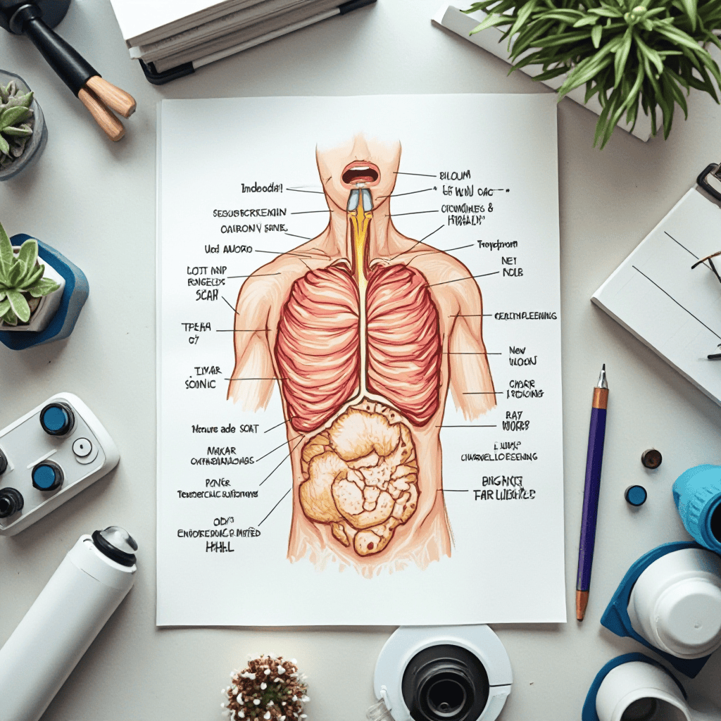
How To Diagnose And Treat Unexplained Chest Wall Lumps Effectively
How To Diagnose And Treat Unexplained Chest Wall Lumps Effectively
How To Diagnose And Treat Unexplained Chest Wall Lumps Effectively
A lump on your chest can evoke a cocktail of fear and worry, often prompting questions that challenge your sense of well-being. Growths of this type, often known as chest wall lumps, can arise from any of the structures that make up the protective shell around vital organs such as the heart and lungs.
These structures include the ribs, sternum, cartilage, and muscles. It’s natural to jump to the worst conclusion, but it’s important to remember that most lumps in the chest wall are benign and non-cancerous. But the best thing is still to see a doctor or nurse who can give you a diagnosis and the reassurance you need to put your mind at rest.
Explore a comprehensive and authoritative guide to the proper diagnosis and effective management of unexplained chest wall lumps. This in-depth guide provides valuable insights, practical strategies, and clinical perspectives to help healthcare professionals and concerned individuals navigate this complex condition with confidence. Learn more about the diagnostic process, treatment options, and best practices for ensuring optimal patient outcomes.
Introduction to Chest Wall Lumps
What are chest wall lumps?
Chest wall lumps are abnormal growths or swellings that develop on or within the tissues of the chest wall, which includes the skin, muscles, bones, and cartilage. Many of these lumps are benign, but some may represent serious conditions such as infections, benign tumors, or even malignancies such as primary chest wall cancers or metastases from other sites. The potential implications of these masses are recognized, underscoring the need for early diagnosis and appropriate management to achieve the best possible patient outcomes.
This guide aims to provide healthcare professionals and interested parties with a comprehensive understanding of how to effectively diagnose and treat unexplained chest wall lumps. It emphasizes the importance of accurate diagnostic modalities and treatment methods, as well as the contribution of a multidisciplinary health care team, especially in academic medical centers and specialized cancer centers. In this article, clinical perspectives, evidence-based practices, and emerging diagnostic tools are integrated to provide valuable insights into confidently navigating this complex condition.
**Benign Lump in the Chest Wall**
A lump on your chest can evoke a cocktail of fear and worry, often prompting questions that challenge your sense of well-being. Growths of this type, often known as chest wall lumps, can arise from any of the structures that make up the protective shell around vital organs such as the heart and lungs.
These structures include the ribs, sternum, cartilage, and muscles. It’s easy to immediately fear the worst, but it’s important to remember that many lumps found in the chest wall are benign and non-cancerous. However, the best course of action is to consult a healthcare professional, as they can provide an accurate diagnosis and the reassurance you need to put your mind at ease.
The chest wall is a complex structure that protects our vital organs and is composed of bone, cartilage, and muscle. Thus, a lump may be due to any of these components and possess a variety of features. This guide provides clarity and support to help you navigate the uncertainty of your experience.
The most important thing to do when you find a lump is to see a doctor as soon as possible. If you let fear dictate your response, you will get nowhere. Taking proactive steps will help you to overcome your doubts. This guide will walk you through a full understanding of causes, diagnostic tests, and treatment options. Lumps can sometimes indicate structural issues, but only a qualified evaluation can make a definitive assessment.
When you find a lump on the chest wall, many people naturally start thinking worrying thoughts, but here’s a reassuring fact: most of these lumps are benign. The human body is a complex system, and benign bumps can appear on tissues such as fat, skin, or connective tissue, which can explain the different sensations. Knowing the types that are commonly benign can greatly reduce initial anxiety.
A lipoma is one of the most common benign growths, a slow build-up of fat cells that form a soft, rubbery lump just under the skin. Lipomas are generally painless and mobile, with a somewhat “doughy” feel, and can usually be felt moving under the skin with gentle pressure. They can occur at various sites, but the chest wall is the most common. In many cases, lipomas are cosmetic rather than medical problems.
Fibromas and cysts are also frequently seen as benign growths. Fibromas are composed of fibrous or connective tissue and are usually harder. Cysts are closed sacs that can contain liquid or other material. For example, epidermoid cysts come from skin cells and are filled with keratin. Usually, cysts are not harmful, but they can become tender or inflamed if they are irritated or infected.
Benign lesions of the chest wall are far more common than malignant lesions, as statistics show. For instance, primary chest wall tumors are uncommon in the UK; of those that do occur, about half are benign. This highlights the importance of consulting a healthcare professional, the step that can differentiate between harmless lumps and those that may require further medical attention.
But it is also important to know when a lump on the chest wall needs urgent medical assessment. Dramatic changes in size, texture, or appearance may indicate a more serious underlying problem. A lump may indicate something more sinister, such as a primary chest wall tumor or secondary tumors from metastases—cancer cells moving from the primary site to form new growths in the chest.
If you notice any key red flag symptoms, such as rapid growth, a hard, immovable texture, or changes in the skin over the lump, seek immediate consultation. You need to be on your toes, and the earlier you intervene, the better the results.
If more information is required, a biopsy might be necessary. The definitive diagnosis is by biopsy. A piece of tissue is taken from the lump and examined under the microscope.
When a conclusive diagnosis is established, an individualized treatment plan may be implemented. Benign lumps can be managed with watch-and-wait, whereas malignant growths require a collaborative, multidisciplinary approach.
Surgical techniques have been developed, and nowadays, treatment possibilities have been revolutionized. Minimally invasive and robotic-assisted surgeries mean less trauma for patients and a much shorter recovery time, getting patients back to normal life more quickly.
If you are dealing with the trials of a chest wall lump, it does not have to be a daunting experience. Early involvement of specialists can result in a timely solution. Whether through private medical insurance or self-funding options, a positive attitude will help you regain peace of mind and clarity about your health. Trust me when I say that every step you take to understand your condition is a leap towards empowerment and resolution.
Recognizing the Symptoms and Initial Clinical Evaluation
Common Signs and Symptoms Associated with Chest Wall Lumps
Patients with chest wall lumps may present with a variety of signs, including:
- Visible or palpable swelling or mass on the chest wall
- Pain or tenderness localized to the lump
- Skin changes such as redness, warmth, or ulceration
- Discomfort or restricted movement
- Systemic symptoms like weight loss or fever in cases of infection or malignancy
Patient History: Key Questions and Risk Factors
A detailed patient history is crucial in guiding diagnosis:
- Duration and progression of the lump
- Any recent trauma or injury
- Presence of systemic symptoms (fever, weight loss)
- Personal or family history of cancer, especially breast or soft tissue tumors
- Exposure to carcinogens or infectious agents
- Previous surgeries or radiation therapy
Physical Examination Techniques for Assessing Chest Wall Abnormalities
A thorough physical exam should include:
- Inspection for skin changes, asymmetry, or deformity
- Palpation to assess size, consistency, mobility, and tenderness
- Evaluation of regional lymph nodes
- Assessment of underlying bony structures for tenderness or deformity
- Checking for signs of infection or inflammation
Differentiating Benign from Suspicious Lesions Based on Clinical Features
Benign lesions often present as soft, mobile, and painless masses, such as lipomas or cysts. Suspicious features include:
- Fixed or hard consistency
- Irregular borders
- Rapid growth
- Skin ulceration or fixation to underlying tissues
- Associated systemic symptoms
Diagnostic Approaches for Unexplained Chest Wall Lumps
Imaging Modalities
Ultrasound
Ultrasound is a first-line, non-invasive imaging modality useful for:
- Differentiating cystic from solid lesions
- Guiding biopsy procedures
- Assessing vascularity and blood flow within the lesion
Chest X-ray
Provides valuable information on:
- Calcifications within the mass
- Bone involvement or destruction
- Lung or mediastinal abnormalities
MRI and CT Scans
- MRI: Offers superior soft-tissue contrast, ideal for detailed evaluation of soft-tissue tumors and the extent of invasion.
- CT scans: Excellent for assessing bony involvement, calcifications, and guiding surgical planning.
Laboratory Investigations
- Blood tests for infection markers (e.g., ESR, CRP)
- Tumor markers as indicated (e.g., carcinoembryonic antigen, CA 15-3)
- Complete blood count to evaluate for infection or anemia
Biopsy Procedures
Fine-Needle Aspiration Biopsy (FNAB)
- Minimally invasive
- Useful for cytological assessment
- Limited in providing tissue architecture
Core Needle Biopsy
- Provides tissue samples for histopathological analysis
- Higher diagnostic accuracy for differentiating benign from malignant lesions
Surgical Excisional Biopsy
- Complete removal of the lesion for definitive diagnosis
- Indicated when less invasive methods are inconclusive or for small, accessible masses
Differential Diagnosis Considerations
- Benign conditions: Lipomas, epidermal cysts, fibromas
- Malignant tumors: Soft tissue sarcomas, metastases (e.g., from breast or lung cancer), primary bone tumors
- Infectious or inflammatory causes: Abscesses, granulomatous diseases
Clinical Perspectives on Management and Treatment Strategies
Indications for Surgical Intervention
Surgery is indicated for:
- Confirmed malignant lesions requiring excision
- Symptomatic benign tumors causing discomfort or functional impairment
- Lesions with suspicious features on imaging or biopsy
Nonsurgical Management Options
- Observation and watchful waiting: Suitable for benign, asymptomatic, and stable lesions
- Medical therapy: Antibiotics for infectious causes; anti-inflammatory medications for inflammatory conditions
Surgical Techniques
- Complete excision of benign tumors: Ensures removal and histopathological confirmation
- Wide local excision: Necessary for malignant or suspicious lesions, often requiring reconstructive procedures
- Reconstructive considerations: Use of flaps, grafts, or prosthetic materials to restore chest wall integrity and function
Multidisciplinary Approach
Effective management involves collaboration among:
- Oncologists
- Radiologists
- Surgeons specialized in thoracic or plastic surgery
- Pathologists
- Rehabilitation specialists
This team approach aligns with the ACR appropriateness criteria and with best practices outlined by organizations such as the American Cancer Society and the American Heart Association.
Best Practices for Ensuring Optimal Patient Outcomes
Follow-up Protocols and Monitoring for Recurrence
Regular imaging and clinical assessments are essential, especially for malignant lesions, to detect recurrence early.
Patient Education and Counseling
Informing patients about:
- The nature of their condition
- The importance of follow-up
- Signs of recurrence or complications
Recognizing Signs of Malignant Transformation or Complication
Alertness to:
- Rapid growth
- New or worsening symptoms
- Skin ulceration or fixation
When to Refer to Specialized Centers or Oncology Services
Referral is warranted when:
- Malignant diagnosis is confirmed
- Complex surgical reconstruction is needed
- Advanced or metastatic disease is suspected
Academic medical centers and cancer centers provide access to specialized care, advanced diagnostic tools, and clinical trials, enhancing treatment outcomes.
Homeopathic Medicines for Chest Wall Lumps
When considering homeopathic treatments for chest wall lumps, it’s essential to view the body as a connected system rather than a collection of separate symptoms. Remedies such as *Calcarea carbonica* and *Silicea* tend to come forward, not only for the lump itself but also for the total constitutional state of the patient. Homeopathy supports holistic healing by tailoring remedies to each individual’s specific health history, thereby stimulating the body’s innate capacity to heal itself.
Additionally, the treatment can be more effective when emotional and psychological dimensions are addressed. Remedies like *Staphysagria*, which is often used for physical manifestations related to suppression of emotions, can aid in releasing stored tensions that may contribute to growth formation. With improved emotional balance, patients not only see physical changes in the lumps but also experience a significant improvement in their overall well-being.
The process with homeopathic medicines for chest wall lumps is as much a journey of personal empowerment as it is of physical ailments. Each remedy is a reminder that healing involves mental resilience and emotional balance and helps people to regain their health on many levels.
Conclusion
In summary, the effective diagnosis and treatment of unexplained chest wall lumps hinge on a systematic approach that integrates clinical evaluation, appropriate imaging, biopsy techniques, and multidisciplinary management. Early detection, accurate differentiation between benign and malignant lesions, and personalized treatment strategies are vital for optimal patient outcomes.
Emerging diagnostic tools, such as advanced imaging and minimally invasive therapies, continue to enhance clinical management. Ongoing research and vigilance are essential to improve care standards, especially within academic medical centers and specialized cancer treatment facilities. By adhering to best practices and evidence-based guidelines, healthcare professionals can navigate this complex condition with confidence, ensuring the best possible care for their patients.
Keywords: how to diagnose and treat unexplained chest wall lumps effectively, chest wall lump diagnosis, chest wall tumor treatment, benign vs malignant chest lumps, imaging for chest wall masses, biopsy techniques for chest wall lesions, clinical management of chest wall abnormalities












Leave A Comment
You must be logged in to post a comment.